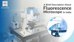
Fluorescence microscopy is an essential tool for studying cell physiology. Fluorescence microscopy is a type of microscope that studies the properties of organic and inorganic materials by using fluorescence and phosphorescence.
Fluorescence is the emission of light by a substance that has absorbed light or other electromagnetic energy while phosphorescence is a kind of optical property that is related to fluorescence.
Microscopy Is a Basic Research Tool
Microscopes are significant research and discovery tools that have helped in the discovery of numerous ground-breaking discoveries over the years. Fluorescence microscopy integrates the magnifying properties of a light microscope with fluorescence technology, which allows the activation of fluorophores – fluorescent chemical substances and the measurement of their emissions.
Fluorescence microscope in India allows scientists to observe where specific cell types or chemicals are present inside tissues or cells.
Components of Fluorescence Microscopy
- Fluorescent dyes (Fluorophore)
- A light source
- The excitation filter
- The dichroic mirror
Applications:
Fluorescence microscopy is used to detect various structures, chemicals, or proteins inside the cell. Almost every component of living or fixed cells and tissues can be labelled and examined using the technique at high magnifications.
When it comes to fluorescence microscopy, the fluorophore can be just as important as the microscope itself, and the type of fluorophore photographed affects the excitation wavelength and detection wavelength. The excitation wavelengths contain a small range of energy that the fluorophore can absorb and cause it to transition to an active state.
Once stimulated, a wide range of excited, a variety of emissions or transitions back to a lower energy state is prepared, leading to an emission spectrum.
Fluorescence Microscopy for Cell Labeling
Fluorophores, such as fluorescent dyes, are used to label cell structures, significantly increasing the visible qualities of biological or chemical structures. These can be modified or combined to have specific target ability in medical applications. High-specificity fluorophores are commonly used in medical diagnostics, and new research is finding new applications for them.
Fluorescence Microscopy for Protein Characterization
Fluorescence microscopy can be used in a variety of experiments that use different types of fluorophores. The detection of proteins that have been labeled with antibodies that are attached to or “conjugated” to fluorescent compounds is one of the most common applications of fluorescence microscopy. Intracellular dynamic protein interactions are important for a wide range of biochemical functions.
We are the best fit for you if you are looking for Fluorescence Microscopy in India
Quasmo India Microscope trades in several different microscopes. We offer Fluorescent Research Microscopes PZQ-102, PZQ-106, and Fluorescent Research Microscopes QX-50, Star- 7FL, and Malaria Detection Microscope PZQ-101 that are the best categories of our Fluorescence Microscopy in India.
In the Ultraviolet wavelength range, we provide a variety of continuously variable filters capable of examining the spectral properties of a specific sample with exceptional transmission values and deep blocking. If you would like any more information about our products, you can contact us, we always available to assist you.
