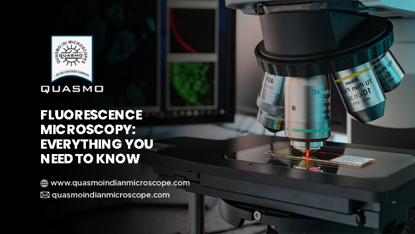
If we start at the beginning and talk about what a fluorescence microscope is and what it does, we can say that it is an optical microscope that studies the properties of organic and non-organic objects using fluorescence and phosphorescence instead of or in addition to reflection or absorption.
The light emitted by a substance with absorbed light or another electromagnetic radiation is known as fluorescence.
When we discuss what phosphorescence is, we can say that it is a sort of photo-luminescence similar to fluorescence.
In the early twentieth century, the fluorescence microscope was invented. This incredible invention is performing admirably.
Fluorescence microscopy is a powerful analytical tool that combines light microscopy’s magnifying properties with fluorescence visibility. It is a phenomenon in which a fluorescent molecule called a fluoroscope absorbs and emits a small range of light wavelengths. This microscopy is performed by combining a bright light source, specific filters, and a method of fluorescent marking material with a basic light microscope.
What is the underlying principle?
This procedure is based on a simple idea. When light reaches the arc lamp, it is directed towards the exciter filter, which determines the excavation length.
Structure and Working
The fluorescent microscope’s principal components are quite similar to those of a standard light microscope. The type of light source and the employment of specific filter components are the two key variations.
The light source lamp, excitation filter, dichroic mirror, and emission filter are all standard components of this microscope. Getting this top-notch source from the leading microscope manufacturer in Ambala is a piece of cake.
Through the objective lens, the light of the excitation wavelength is focused on the specimen. The objective focuses on the fluorescence emitted by the specimen on the detector. Only reflected excitation light and the emitted light reach the aim since most of the excitation light is transmitted through the sample.
What Are The Applications of This Microscope?
This microscope is handy for spotting features in both preserved and living biological samples.
Today, this microscope is standard equipment for scientific research and processes, and this incredible discovery is changing the face of science. The fluorescence microscope in India has been steadily expanding in popularity owing to the growing demand for this technology. The reason for its ever-increasing magnitude is the quality of service provided by this source. It allows for multi color staining, structural labeling, and the measurement of a cell’s physiological condition.
Advantages
Fluorescence microscopy is one of the most popular technologies for live-cell observation and elucidating the structure of bio molecules in tissues and cells. It allows them to be studied without the need for hazardous and time-consuming staining processes. Cell dynamics can be examined in living samples with a precision and sensitivity capable of tracking the path of a single protein throughout its lifetime, or samples can be fixed before the addition of a fluoroscope, halting the metabolism of cells at a point in time and allowing detailed measurements of the preserved specimen.
When employed in conjunction with con focal microscopy, the great degree of specificity provided by contemporary fluoroscope probes allows specific proteins or other biological components to be stressed and highlighted. This results in a clear three-dimensional internal structure of the cell. Multiple overlapping systems can be discovered simultaneously within the same sample due to the diversity and specificity of the available probes.
