Author Archives: qmadmino

Fluorescence microscope

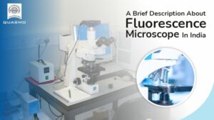
If we start at the beginning and talk about what a fluorescence microscope is and what it does, we can say that it is an optical microscope that studies the properties of organic and non-organic objects using fluorescence and phosphorescence instead of or in addition to reflection or absorption.

The light emitted by a substance with absorbed light or another electromagnetic radiation is known as fluorescence.

When we discuss what phosphorescence is, we can say that it is a sort of photo-luminescence similar to fluorescence.

In the early twentieth century, the fluorescence microscope was invented. This incredible invention is performing admirably.

Fluorescence microscopy is a powerful analytical tool that combines light microscopy’s magnifying properties with fluorescence visibility. It is a phenomenon in which a fluorescent molecule called a fluoroscope absorbs and emits a small range of light wavelengths. This microscopy is performed by combining a bright light source, specific filters, and a method of fluorescent marking material with a basic light microscope.

What is the underlying principle?

This procedure is based on a simple idea. When light reaches the arc lamp, it is directed towards the exciter filter, which determines the excavation length.

Structure and Working

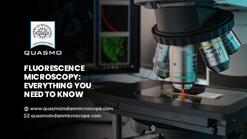
The fluorescent microscope’s principal components are quite similar to those of a standard light microscope. The type of light source and the employment of specific filter components are the two key variations.

The light source lamp, excitation filter, dichroic mirror, and emission filter are all standard components of this microscope. Getting this top-notch source from the leading microscope manufacturer in Ambala is a piece of cake.

Through the objective lens, the light of the excitation wavelength is focused on the specimen. The objective focuses on the fluorescence emitted by the specimen on the detector. Only reflected excitation light and the emitted light reach the aim since most of the excitation light is transmitted through the sample.

What Are The Applications of This Microscope?

This microscope is handy for spotting features in both preserved and living biological samples.

Today, this microscope is standard equipment for scientific research and processes, and this incredible discovery is changing the face of science. The fluorescence microscope in India has been steadily expanding in popularity owing to the growing demand for this technology. The reason for its ever-increasing magnitude is the quality of service provided by this source. It allows for multi color staining, structural labeling, and the measurement of a cell’s physiological condition.

Advantages

Fluorescence microscopy is one of the most popular technologies for live-cell observation and elucidating the structure of bio molecules in tissues and cells. It allows them to be studied without the need for hazardous and time-consuming staining processes. Cell dynamics can be examined in living samples with a precision and sensitivity capable of tracking the path of a single protein throughout its lifetime, or samples can be fixed before the addition of a fluoroscope, halting the metabolism of cells at a point in time and allowing detailed measurements of the preserved specimen.

When employed in conjunction with con focal microscopy, the great degree of specificity provided by contemporary fluoroscope probes allows specific proteins or other biological components to be stressed and highlighted. This results in a clear three-dimensional internal structure of the cell. Multiple overlapping systems can be discovered simultaneously within the same sample due to the diversity and specificity of the available probes.

Microscopes have long been used in scientific and medical disciplines. This incredible discovery has made it easier for humans to study even the tiniest of details with extreme care. This great creation has been around for a long time and is still used for study and research. Microscopes have a wide range of uses, from typical school labs to high-tech scientific sectors. And it is practically difficult to execute any scientific activity without them. This vast range of amenities has increased the potential for the best microscope exporters to Germany to expand their firm. Explore new avenues for success.

When it comes to using microscopes, there are a few things to remember. Let’s look at some of the things you should and shouldn’t do when using a microscope.

What Should You Do When Using a Microscope?

1. Slide handling: When using a microscope, slides are like the fundamental plates on which the elements are held. This must be carried out with extreme caution and cleanliness. Before completing any experiment or observation, you should thoroughly clean your hands. As finger oil can cause a cloudy vision and make it impossible to see the required organism.

2. Cleaning with extreme caution: If you want to clean the eyepiece, you shouldn’t rush through it. Breathe into the eyepiece to dislodge the debris, then gently wipe it away with condensed water and lens paper to restore vision clarity.

3. Careful oil cleansing: If you notice any oil on objects, try removing it by dampening a piece of paper with the ethyl ether solution and slowly rubbing the wet paper across the front surface of the lens. Microscope lenses are sensitive, and you should take special precautions to avoid dusting and oiling their surfaces.

Don’ts: When Using a Microscope, Avoid These Mistakes

1. Cosmetics are to be avoided at all costs: There are a few things to consider if you are obliged to work in a scientific laboratory for longer hours. And must use a microscope frequently. Wearing mascara or kajal in your eyes might cause vision problems and cause the lens to cling to the surface. You must aim for flawless lens clarity.

2. Using dry paper to touch the lens: this should be emphasized more. Always make an effort to touch the lens with clean, clear lens paper. You shouldn’t try to touch it with dry paper, and you should be careful about how you hold it. Apart from that, you should store or place the microscope in a secure location away from potential oil or water spills. You should take great care of it and use it as efficiently as possible to extend its life.

3. Rub the lens unnecessarily: You should never rub the lens while cleaning or holding it. It causes hazy sight, and you may not be able to inspect the desired thing adequately. So, whether you’re cleaning or using it, make sure you use only your eyes and not too much of your hands to avoid rubbing or touching it. Cleanliness and clarity are two crucial factors to consider when caring for a microscope lens, and they are their most valuable attributes.

Microscopes have made it easier to pay attention to the finest of details, whether it’s for an observation or a scientific study. Because of its vast range of applications and demands, microscope exporters from India to Germany have been driven to create more and catch the attention of more potential clients to achieve a large market share. Microscopes stand out because of their applications and quality, and it’s all thanks to the brilliant individual who invented them.

The introduction of the microscope simplified things and was a boon to researchers and experimenters who couldn’t afford to postpone their tests or research. This incredible creation occurred millions of years ago. It was once a basic microscope with only one lens. More improvements have led to the availability of the compound microscope, which has two lenses.

Optical microscopes were the first microscopes invented, and they relied on light to observe the sample being examined. The way microscopes work has changed as science has progressed, new procedures have been developed, and new types of microscopes have been discovered. The electron microscope, ultra-microscope, and scanning probe microscopes are examples of innovative inventions in the microscopic realm.

The increased use of microscopes has resulted in an increase in demand for this resource. The most exemplary microscope exporter from India to Saudi Arabia has seen a developing opportunity and gained a competitive advantage over other options. This has accelerated the growth of the industrial sector, and distribution is expanding at a breakneck pace.

Let’s have a look at some of the fascinating facts behind this marvelous invention of humankind.

Amazing Facts About Microscopes-

1. Spherical aberration is the uneven bending of light as it passes through various regions of a lens. Joseph Jackson Lister solved the problem presented by these lenses in 1830. This occurred while he was making a discovery, in which he set lenses at a certain distance apart and realized that the aberration caused by the first lens could be erased.

2. Prior to the invention of microscopes, illnesses were thought to be caused by noxious gases or evil spirits. People began to believe in the existence of germs and funguses after the microscope was established, and they could finally understand what the major causes of sickness are, as well as what it takes to get to the root cause.

3. Microscopes have a long and illustrious history. After the advent of glass in the first century, the Romans began experimenting with various sizes and forms to see objects. Flea glasses were the first microscopes, which were just magnifying glasses with a limited magnification capacity. Flea glasses got their name because they were used to measure microscopic creatures like fleas.

4. Marcello Malpighi is the father of microscopic anatomy. He’s famous for bringing concepts like taste buds and red blood cells to life. He discovered the system of microscopic capillaries connecting the artery and venous networks using a rudimentary early microscope. Thanks to this Italian scientist’s astounding discovery, the relevance of red blood cells and taste buds cannot be neglected by any human today.

5. An electron microscope magnifies images by using electrons rather than light. The electronic transmission microscope, designed by Ernst Ruska in 1931, was the first electron microscope.

6. To see a more extensive range of objects, Robert Hooke used a basic compound microscope. He saw a pore on the cork’s surface while inspecting it but was unaware that he had also discovered plant cells. These pores were later dubbed “pore cells” by him.

Microscope inventions and discoveries have changed the fundamental dynamics of observations, and Microscope exporters to Saudi Arabia and elsewhere have reaped significant benefits in commerce and profit margins. Today’s microscopes have a wide range of applications and are used in a variety of scientific fields.


Laboratory Microscope Suppliers In India

In the technological world, the research field is seeing tremendous growth in recent times. For undergoing biological research, the use of the microscope is inevitable. It ensures to provide a close view of any tiny objects or organisms.

With the help of microscopes, many advancements are happening in the medical field. For finding out if an organism is dangerous to humans, the use of microscopes can help. This equipment is in great demand among the researchers, without which the research field is considered empty. Suppliers are offering products from the microscope manufacturer in Ambala as well to aid the research community. There are many varieties of microscopes with unique advantages.

Diagnose the illness of patients

Microscopes, besides research purposes, aids doctors in identifying the type of infection in the patients. Sometimes it becomes tough to diagnose from the symptoms that patients experience. Dangerous diseases, when left undiagnosed, can lead to the death of the person. Hence the use of microscopes comes into action in these situations. It helps in examining the blood samples of the patients.

When a person is affected due to viruses or bacteria, it can be confirmed if the infection is positive or negative using microscopes. Also, doctors can find the level of infection spread in the body to provide suitable treatments. Buying the equipment from the laboratory microscope suppliers in India can benefit the best diagnosis of diseases. Patients get the best cure for the disease and lead a healthy life.

Aids in the development of vaccines

Microscopes can help to produce the appropriate vaccines for stopping the disease spread. They aid in zooming the virus structure and identify its type and severeness. Hence, researchers can find the equipment to be more beneficial for developing vaccines and manufacturing medicines. Scientists also use this equipment for finding vaccines to prevent disease spread in animals.

Research organisms structure

To understand and realize the structure of tiny organisms, microscopes aid to a great extent. This equipment helps zoom the germs for viewing the organs and their cellular structure. Visualizing these organisms deeper can provide better knowledge about its variant and aid in knowing their benefits and ill effects. Microscopes can offer unique visuals as the organism’s structure cannot be viewed by the naked eye.

Types of microscopes

The microscope comes in different types. The electron microscope can help visualize the organism that is impossible with an optical microscope. A virtual microscope converts the image on the slide as electronic data and facilitates remote visualization. These types can come under industrial, digital, or biological microscopes. Along with this, the microscope camera provides a better view of the objects, specimens, organisms or tissues.

The research field is blooming and offering more medical services to the patients and the medical industry. With the assistance of laboratory microscope suppliers in India, it is possible to distribute the equipment across places. Perform research activities with ease and contribute the best to the wellness of the country. Purchase the best microscope and undergo seamless research with scientists and improve the environmental health as well.


Microscopes have been a major aspect of our science community for over centuries. Scientists and Biologists use microscopes to understand the nature of the universe and tiny living organisms and their functions in the sustainability of life. Over the years, the technology has gone a drastic change from simple dye-based photometry to imaging techniques. And this is why fluorescent microscopes have been invented.

India is a diverse land-based on geography and demography, and this technique can be used to determine the structure of different microorganisms dwelling with us. This eventually helps in understanding their uses and threats. Fluorescence technology has been around for a while, and many firms use it to generate images to a scale of 5 um. But we will not go much into the details. Instead, we will have a simple overview of the technique and understand why we need a fluorescent microscope in India.

Fluorescent microscope – a simple overview

A microscope is an instrument that magnifies tiny objects. Fluorescence microscopy is a technique through which the microscope detects a fluorescence signal, and an image is formed on the windows or monitor to show the labeled molecules. Fluorescence occurs when an electron moves from a lower energy state to a higher energy state. This emits light of a longer wavelength that is then captured by the fluorescent microscope. The detector then creates an image where you can see the samples in magnified form.

Indian context for fluorescent microscope

As already mentioned, India is a diverse country with a different demography. Scientists worldwide have been visiting India for research, and in the late 20th century, they introduced the concept of the fluorescent microscope in India. However, the equipment was highly sophisticated, and the cost of importing was plummeting towards the sky.

But the people in major research fields understood the requirement of such sophisticated equipment for their research. So, they found a way to bring the technology from the international borders to our land. They put their minds into understanding the engineering behind it and soon developed the prototype. This encouraged many people to do engineering on their part and make this equipment suited to the Indian market. Now, you can find many dealers and manufacturers for the fluorescent microscope in India and the products not burning a hole in your pocket. You can find some great microscope manufacturers in Ambala if you are looking forward to buying any microscope.

Applications for the fluorescent microscope in India

Now, we have come to the part where we will understand why we need the fluorescent microscope in India. There is a vast range of applications in the medical field for such instruments. People around the globe use this technique in detecting pathogens, cell functions, and Neuroscience.

The spectral vision imaging in the applications helps the researchers and specialist surgeons to find the root cause of the infectious diseases caused by bacteria, viruses, and other microorganisms. They can also detect the effect of certain chemical compounds on such microorganisms to test their medication effects. This will help in reducing the costs of vaccinations and medicines and effectively treat the disease.


Microscope Exporter From India To Spain

A microscope serves many purposes like tissue analysis, determining the ecosystem’s health, examining forensic attestation, figuring out protein’s role in cells, studying atomic structures, and many more. To get acquainted with the perks and advantages of importing microscopes from India and the factors to consider while selecting the best microscope exporter from India to Spain, read on:

What makes India a great microscope manufacturer and exporter?

If you reside in Spain, importing microscopes from India might be a great option due to the following reasons:

  • Wide variety of microscopes: When it comes to microscopes, one shoe certainly doesn’t fit all sizes. India is a manufacturer and exporter of various types of microscopes. These include biological, metallurgical, polarizing, industrial, and fluorescent, among others. Therefore, no matter what kind of microscope you need, India-based microscope exporters have it all.
  • Cost-effective solution: Since India is a major manufacturer and exporter of microscopes, it has a huge infrastructural base. This means that the microscopes are manufactured at a relatively lower price than they are in other countries. Hence, importing microscopes from India is a more cost-effective solution than it is from any other country.

What factors to consider before deciding on a microscope exporter?

While there isn’t a shortage of microscope exporters to Spain from India, some are better than the rest. Here is a list of factors that you must consider before deciding on an exporter:

  • Quality: Whether the microscopes continuously undergo quality tests or not is also an important factor worth considering. The parameters of product testing must be taken into consideration, too. If the microscopes in which the exporter deals successfully pass all the quality tests, you can surely trust the exporter.
  • Distribution network: The exporter must have access to a well-established distribution network. The lack of a stable microscope distribution network will result in postponement in deliveries. To ensure quick delivery of your ordered microscopes, trust only the exporter that boasts of an efficient distribution network.
  • Pricing: The perkiest benefit of having several microscope exporters is that you can always pick and choose. Compare and contrast the prices before making the final decision. Some exporters deliver quality microscopes at competitive prices, which lend them a competitive edge over their contemporaries. One must resort to such exporters.
  • Packaging: Nobody would want to receive damaged products. Microscopes are fragile; hence, they must be carefully packed. Ensure that the microscope exporter guarantees the use of sturdy packaging.
  • Other factors: Other significant factors such as experience obtained in the field, responsiveness to queries, and utilization of the latest technology in the manufacturing procedure must also be considered before finalizing the suitable microscope exporter.

Thus, India serves as a quality exporter of microscopes to Spain and other parts. However, one must not forget to consider certain factors like the ones mentioned by us before finalizing the exporter.


Microscope Exporter From India To The USA

The microscope was an important invention of the 16th century that allowed us to enter the world of microorganisms. Over time, people have used high-power lenses to unravel the world of microbes, and this has helped humanity understand key features of the structures in the smallest world. Now, this aided in developing medicines and drugs to avoid the harmful germs in doing the damage. However, making the microscopes and delivery has been crucial from the commercial perspective.

The USA has become one of the prevalent countries to invest its resources in biology and developing medications. But all the equipment costs are sky-rocketing. This is why every university and research facilities are relying on exporting major equipment from outside. Being one of them, Microscope is also being imported to the United States of America from different countries. So, here we will discuss the major benefits derived from microscope exporters to the USA.

Microscope exporter from India to the USA – the benefits

There is more than one benefit in connecting with a microscope exporter from India to the USA. And we are not talking only about the commercial benefits. Knowledge transfer has always been an eminent part of cultural society. Moreover, we live in a society where knowledge has overcome international boundaries to bring together humanity. So, the following are a few benefits that the faculties can reap from microscope exporters to the USA:

  • Evolutionary microscopes –

Don’t get the term wrong, as it may sound like a microscope designed to understand evolution. Well, in a way, it is the case, but that is not entirely the one. Evolutionary microscope encompasses the variations in microscope based on their applications. So, you don’t have a microscope to study tiny organisms or cells. Instead, you can now use microscopes to study atoms, metallurgical components, zoom microscopes, surgeries, and many more.

  • Cost curtailing –

The major financial benefit of importing costly equipment like microscopes is that you can get a high-resolution microscope at an affordable price if you import from India or its neighbouring countries.

  • Image enhancement –

Many ultra-resolution microscopes are available with the exporters that need an HDMI connection to connect through a monitor to study anything placed underneath the microscope. Searching for the same in the indigenous market may be time-consuming. The exporters know the needs, and they provide a wholesome solution.

  • Low duties and taxes –

Importing and exporting criteria are different for different countries. Therefore, while you may experience low duties and taxes while importing from one country, the duties may rise unexpectedly for other countries. Thankfully, due to the international commercial laws, the import duty of the microscopes is less from India. Thus, it adds to the cost-benefit.

  • Quality –

Quality is a major factor for this high-cost equipment. Therefore, it is very important to maintain quality. But this raises a question as to how microscope exporters to the USA can have competitive pricing and still maintain quality. This is done so as the labour cost is lower. So, they can spend a proportionate budget on material resources than labour and offer quality products at an affordable price.

Now that we have established the major benefits of importing microscopes, you must also head out to different exporters and compare their proposals.


Fluorescence Microscope In India

Fluorescence microscopy is an essential tool for studying cell physiology. Fluorescence microscopy is a type of microscope that studies the properties of organic and inorganic materials by using fluorescence and phosphorescence.

Fluorescence is the emission of light by a substance that has absorbed light or other electromagnetic energy while phosphorescence is a kind of optical property that is related to fluorescence.

Microscopy Is a Basic Research Tool

Microscopes are significant research and discovery tools that have helped in the discovery of numerous ground-breaking discoveries over the years. Fluorescence microscopy integrates the magnifying properties of a light microscope with fluorescence technology, which allows the activation of fluorophores – fluorescent chemical substances and the measurement of their emissions.

Fluorescence microscope in India allows scientists to observe where specific cell types or chemicals are present inside tissues or cells.

Components of Fluorescence Microscopy

  • Fluorescent dyes (Fluorophore)
  • A light source
  • The excitation filter
  • The dichroic mirror

Applications:

Fluorescence microscopy is used to detect various structures, chemicals, or proteins inside the cell. Almost every component of living or fixed cells and tissues can be labelled and examined using the technique at high magnifications.

When it comes to fluorescence microscopy, the fluorophore can be just as important as the microscope itself, and the type of fluorophore photographed affects the excitation wavelength and detection wavelength. The excitation wavelengths contain a small range of energy that the fluorophore can absorb and cause it to transition to an active state.

Once stimulated, a wide range of excited, a variety of emissions or transitions back to a lower energy state is prepared, leading to an emission spectrum.

Fluorescence Microscopy for Cell Labeling

Fluorophores, such as fluorescent dyes, are used to label cell structures, significantly increasing the visible qualities of biological or chemical structures. These can be modified or combined to have specific target ability in medical applications. High-specificity fluorophores are commonly used in medical diagnostics, and new research is finding new applications for them.

Fluorescence Microscopy for Protein Characterization

Fluorescence microscopy can be used in a variety of experiments that use different types of fluorophores. The detection of proteins that have been labeled with antibodies that are attached to or “conjugated” to fluorescent compounds is one of the most common applications of fluorescence microscopy. Intracellular dynamic protein interactions are important for a wide range of biochemical functions.

 We are the best fit for you if you are looking for Fluorescence Microscopy in India

Quasmo India Microscope trades in several different microscopes. We offer Fluorescent Research Microscopes PZQ-102, PZQ-106, and Fluorescent Research Microscopes QX-50, Star- 7FL, and Malaria Detection Microscope PZQ-101 that are the best categories of our Fluorescence Microscopy in India.

In the Ultraviolet wavelength range, we provide a variety of continuously variable filters capable of examining the spectral properties of a specific sample with exceptional transmission values and deep blocking. If you would like any more information about our products, you can contact us, we always available to assist you.


Surgical microscope manufacturer in india

The surgical microscope is an electronic device that doctors use in surgery. Nowadays, the surgical microscope is come up with their unique feature of magnifier as it also used to see tissues largely. A surgical microscope is of great use for doctors and patients because of its exceptional features. Moreover, its usage is long-lasting as it is made up of good quality raw material. It is also available in warranty.

A Surgical microscope manufacturer in india in large quantities. The main advantages are it is not a lengthy and difficult procedure as the incision is small. The surgical process takes less time. Its usage is worldwide like investigations. It is also available as waterproof. It could be used as a nanodevice used by doctors for detecting eye numbers. They are compatible with IOLs which results in better vision. The incision is small, which is beneficial for patients who have diabetes or with other comorbidities.

 More About Surgical Microscope

  • A surgical microscope is used in biological labs for observing cells and tissues.
  • A surgical microscope could be used as a nanodevice used in biological labs, useful during investigations.
  • It is useful to patients whose corneal has been compromised and have a zonular weakness as it uses less phaco energy.
  • The surgery doesn’t injections filled with anaesthesia which is used for unconscious patients.

Features Of Microscope

Quasmo Indian Microscope is the best Surgical microscope manufacturer in india, it is used by doctors to observe cells, tissues, and other biological specimens.

  • The microscope is used for recovering many diseases like diabetes, eye cataract.
  • Its usage is worldwide for investigations, detecting diseases. It is also available as waterproof
  • Many patients can avail of this machine at home as it is easily affordable and less expensive.
  • The success of each microscope depends upon the professional maturity of experts.

There are many surgical microscopes manufactured in India which experts personally test.

Winding Up

The surgical microscope is a magical instrument used by doctors in biological labs to detect diseases, observe cells and tissues. It is also used in the investigation during murders. The main advantages of the surgical microscope are it is not a lengthy and difficult procedure as the incision is small. The surgery doesn’t require injections filled with anaesthesia used for unconscious patients so that they don’t feel any pain during surgery. A surgical microscope is useful to patients whose corneal has been compromised and have a zonular weakness as it uses less phaco energy.

They are compatible with IOLs which results in better vision. The incision is small, which is beneficial for patients who have diabetes or with other comorbidities. Surgical microscope has such amazing features which are acceptable and popular among doctors and patients. These are the best microscope in the world which give doctors magical powers for recovering patients fast. It also maintains a good relationship between doctors and patients.

Malaria Detection Microscope

Benefits of Microscope in Malaria Diagnosis:

Malaria Detection Microscope

A quote “Health is Wealth” is maybe a clichéd quote yet very true!  In this particular article, we will look at what are the benefits of Malaria Detection Microscope that can be leveraged for detecting malaria, a deadly disease, and the commitment of building a low-cost, effective and open-source solution.

Abstract

Malaria is a fatal, communicable mosquito-borne disease produced by Plasmodium parasites. These parasites are spread by the stings of infected female Anopheles mosquitoes.

Malaria is still today considered as one of the highest killer and serious parasitic diseases striking tropical countries with major challenges. It is an overwhelming problem in developing countries in the tropical region. Most deaths occur in Africa, where a child dies around every minute from malaria, and where malaria is a leading cause of childhood neuro-disability. According to the World Malaria Report 2016, an estimated 3.2 billion people in 95 countries and territories are at risk of being infected with malaria and developing the disease, and 1.2 billion are at high risk.

The World Health Organization recommends that malaria treatment be based on demonstration of the infecting Plasmodium parasite species. The discovery of Plasmodium parasites by light microscopy is still the chief method of malaria diagnosis in most health care facilities throughout the world.

Currently, in the northeastern parts of India, malaria case detection, especially in remote areas, depends heavily on the microscopy method and it is being regarded as one of the gold standard methods for malaria diagnosis towards the identification of Plasmodium.

At community points of care, Malaria rapid diagnostic tests (RDTs) are recommended as they are accurate and rapid and have the potential to significantly improve and standardize malaria diagnosis.

Let’s Chalk Down The Symptoms Of Malaria:

Clinical diagnosis is the least expensive; the most commonly used mode of malaria diagnosis and is the reason for self-treatment. However, the overlap of malaria symptoms with other tropical diseases and can result in misdiagnosis. Most often the first symptoms of malaria are fever, chills, sweats, headaches, muscle pains, nausea, and vomiting. Along with that there are also found in other diseases such as the “flu” and common viral infections, elevated temperature, perspiration, tiredness. Confusion, coma, neurologic focal signs, severe anemia, respiratory difficulties are also more striking and may increase the index of suspicion for malaria.

Benefits of the microscope in Malaria Diagnosis

Microscopy is an equipped, comparatively simple technique that is commonly used in laboratories. Any laboratory that can conduct regular hematology tests that are implemented to perform a thin and thick malaria smear. A microscopy test provides valuable information within a few hours of collecting the blood. Firstly, it is determined that malaria parasites are present in the patient’s blood. Once the diagnosis is confirmed – normally by detecting parasites in the thick smear – the laboratorial examines the thin smear to fix the malaria species and the parasitemia. The thin and thick smears can provide vital information to the doctor to guide the initial treatment decisions that need to be made acutely.

Benefits of the microscope in Malaria Diagnosis: Microscopic malaria diagnosis prevails in the low direct cost that gives it a distinct advantage in resource-poor settings. Other existing diagnostic methods or any new method have to verify that they can provide similar ease of use.

Important things should be following while using a malaria detection microscope:

Timely and accurate diagnosis of malaria is significant for efficient treatment and to stop transmission in communities. Microscopic tests must be performed quickly when commanded by a health-care provider. Besides, these tests should not be sent out to reference laboratories with results available only days to weeks later. Health-care providers must receive results from these tests within hours to appropriately treat their patients infected with malaria.

End Note:

Due to the huge benefits of the microscope in Malaria Diagnosis, Microscopy performed better and is more reliable than the first response in areas with low parasite density among patients less than 6 years of age. Rapid diagnostic tests could be useful in areas with high parasite density as an alternative to smear microscopy