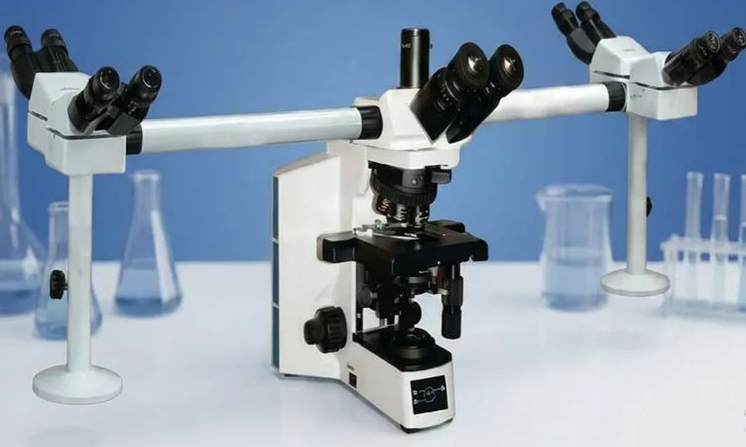
Home • Our Products • Multi Viewing Microscope • Advance Mul Head Microscope Model: PH-999
Multi Viewing Microscope for Tomorrow's Innovators
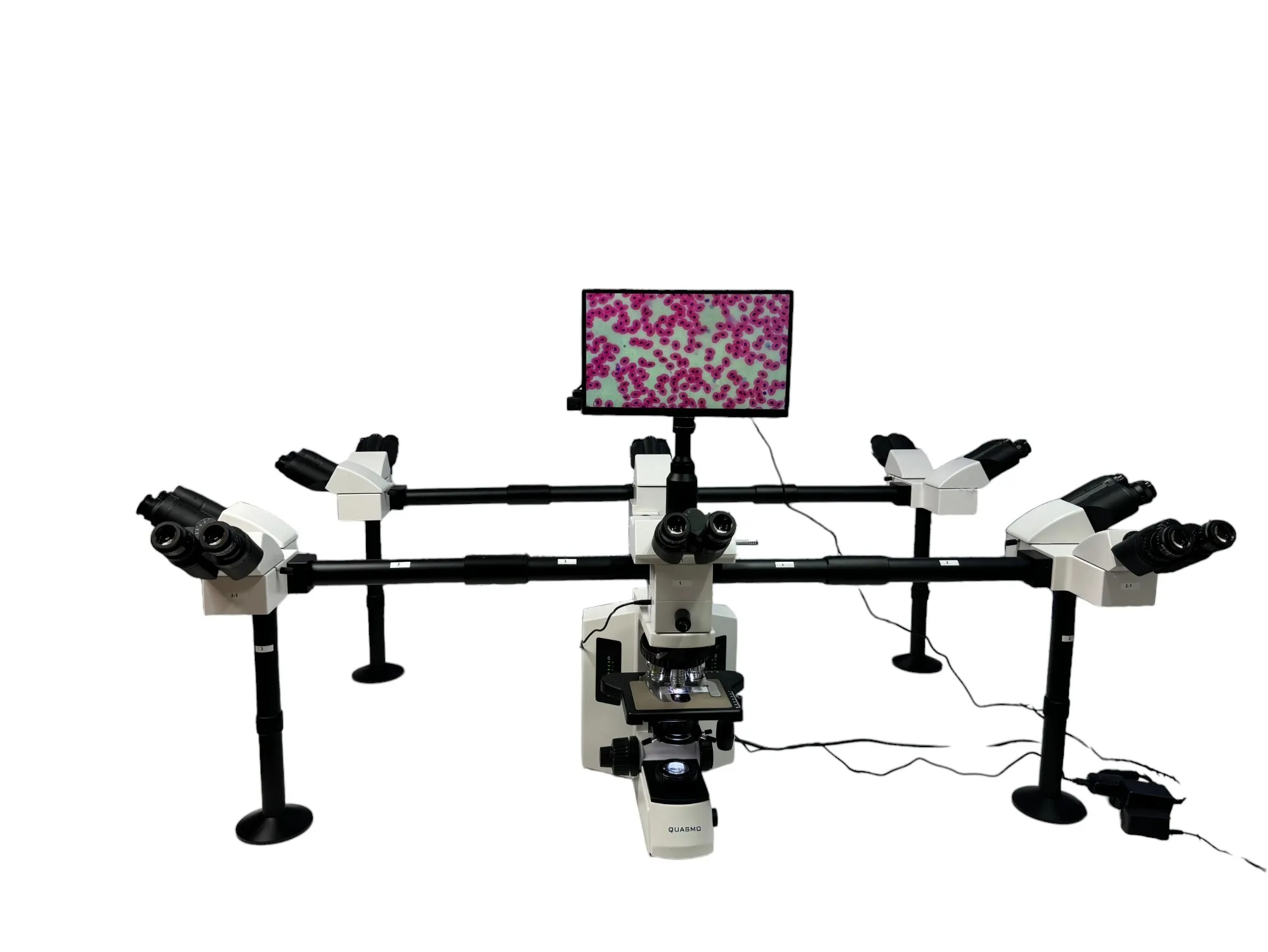
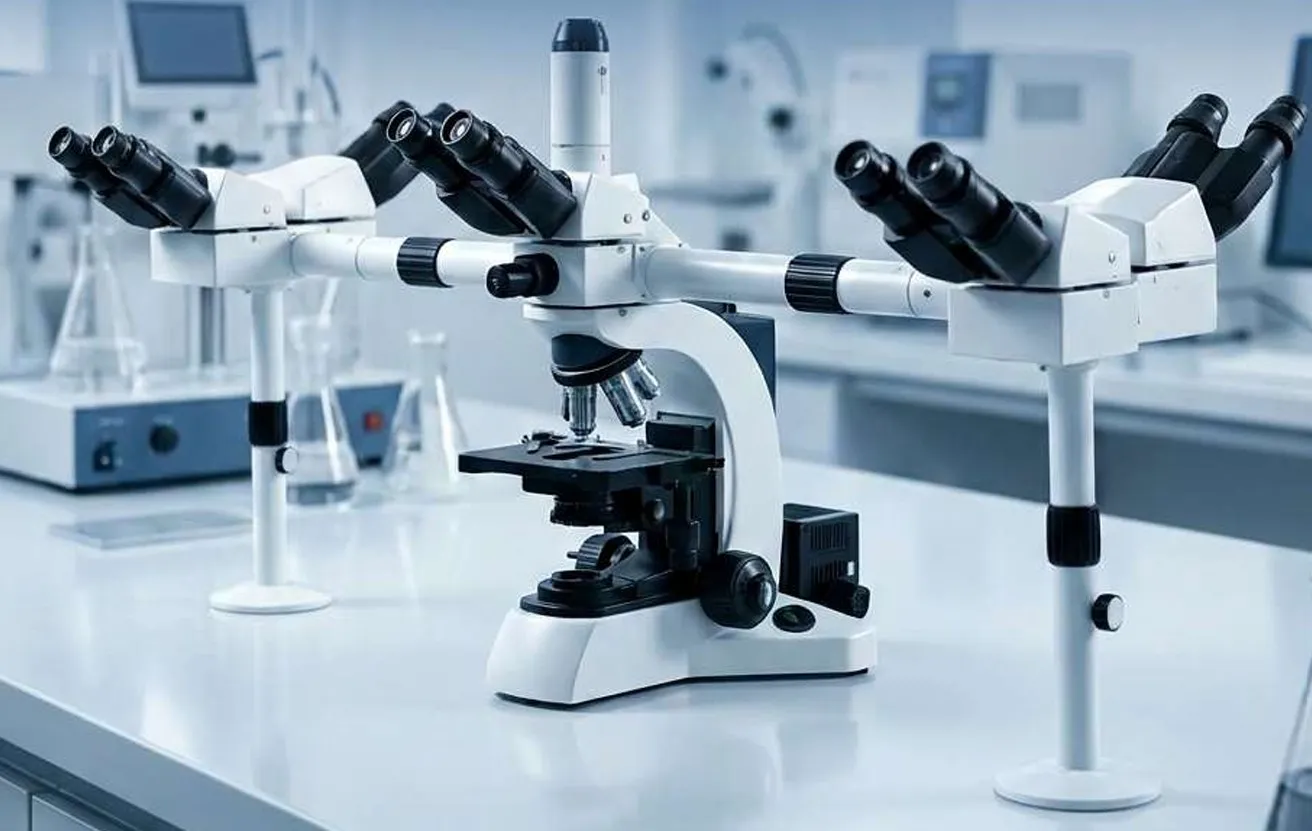
Multi Viewing Microscope is designed to allow simultaneous observation of the same specimen by multiple users. Equipped with two or more viewing heads, it is ideal for teaching, training, clinical discussions, and collaborative analysis. It delivers clear, synchronized images to all viewers, ensuring accurate demonstration and effective knowledge sharing in laboratories and educational institutions.
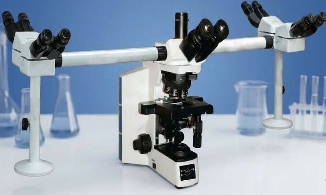
Advance Mul Head Microscope Model: PH-999
Advance Multi Head Microscope – Model: PH-999 is a high-performance microscope designed for teaching, laboratory, and research applications. Its multi-head viewing system allows multiple users to observe specimens simultaneously, delivering clear imaging, precision focusing, and reliable performance for medical and scientific use.
Advance Mul Head Microscope Model: PH-999
Advance Multi Head Microscope – Model: PH-999 is a high-performance microscope designed for teaching, laboratory, and research applications. Its multi-head viewing system allows multiple users to observe specimens simultaneously, delivering clear imaging, precision focusing, and reliable performance for medical and scientific use.
Product Description
The PH-999 Advance Multi Head Microscope is a high-performance, research-grade microscope specially designed for teaching, clinical diagnosis, laboratory observation, and collaborative research. It features a multi-viewing head system that allows multiple users to observe specimens simultaneously, making it ideal for medical colleges, pathology labs, training institutes, and research centers. Equipped with an infinity-corrected optical system, high-resolution objectives, LED illumination, precision focusing
Product Packaging
Thermocol/Styrofoam Packing And Outer Cardboard Box.
Product Specification
| Optical System: | Universal color corrected infinity optical system |
| Stand: | Teaching ten headed microscope designed to enable simultaneous viewing of a specimen by up to ten users. Suitable for medical, paramedical, biological and teaching laboratories. Facilitates guided instruction, coordinated observation, and group discussion through multiple viewing heads. |
| Eyepiece: | Paired wide field eyepieces with 10X magnification. Diopter adjustment on both eyepieces. Field Number (F.N.) 22mm Simultaneous live viewing through eyepiece and monitor display. |
| Teaching Eyepiece: | Wide field 10X magnification, FOV minimum 22 mm, both sides with diopter adjustment. |
| Nosepiece: | Six-position or higher revolving nosepiece. Slot provision for analyzer and DIC slider. Preferable coded nosepiece configuration. |
| Stage: | Mechanical stage with ceramic coating. Minimum stage size: 185 × 135 mm. Double slide holding capacity. Travel range: minimum 76 × 52 mm. |
| Analyzer & Polarizer: | One set of analyzer and polarizer with all standard accessories.20 cc immersion oil included. |
| Focusing : | Coaxial coarse and fine focusing controls. Minimum 25 mm stage stroke. Coarse adjustment limit stopper. Fine focus adjustment grada on of 1 µm or better. Torque adjustment facility for coarse focusing knobs. |
| Objectives: | infinity-corrected objectives an-fungal coated. Objective transmittance: more than 80% from 400–700nm. An-fungus treated optics. Plan Achromat 2X Objective, NA 0.06 Working Distance 30.0mm with unbreakable objective container. Plan Achromat 4×: NA 0.10, Working Distance 18.5 mm with unbreakable container. Plan Achromat 10×: NA 0.25, Working Distance 10.6 mm with unbreakable container. Plan Achromat Phase Contrast 20×: NA 0.40, Working Distance 0.9 mm with unbreakable container. Plan Achromat Spring Loaded Phase Contrast 40X Objective, NA 0.65, WD 0.36 mm. with unbreakable container. Plan Achromat 100× (Oil Immersion): NA 1.40, Working Distance 0.13 mm, spring-loaded with unbreakable container |
| Observation Head: | Wide field ergonomic trinocular observation tube. Continuous tilting angle range from 5° to 35° . Three-way light path distribution: 0 / 50 / 100. Simultaneous viewing and imaging capability. Tube secured with special dovetail mechanism. Minimum field number: 22 mm. Fluorescence upgrade compatability. |
| Multi-Head Teaching System: | Teaching a achment for 1 + 4 users. Ergonomic binocular heads with continuous tilting angle range from 5° to 35°. Paired wide field eyepieces with minimum 10X magnification. Diopter adjustment on both eyepieces. Field Number (F.N.) 22mm. Multicolor LED arrow pointer for teaching and indication. Convenient and sensitive control buttons. |
| Condenser: | Universal condenser with minimum five positions. Numerical Aperture (NA) 0.9. Suitable for Bright Field, Phase Contrast, Polarizing, and Dark field applications. |
| Illumination: | Built-in Koehler illumination system. Ergonomic microscope stand with high-intensity LED illumination. 14 W LED light source. LED life expectancy: 60,000 hours. Illumination intensity equivalent to 12 V / 100 W halogen illumination. Variable light intensity control for brightness adjustment. |
| Camera: | 8 MP Quasmo color CMOS WiFi camera. Sensor size: 1/1.8 inch (7.140 mm × 4.980 mm).Maximum snapshot resolution: 2592 × 1944 pixels. HDMI/WLAN/PC resolution: 1920 × 1080 pixels. Pixel size: 2.4 × 2.4 µm.8-bit A/D converter. Exposure me range: 1 ms – 918 ms. Live frame rates: Up to 30 fps on PC. Up to 60 fps through HDMI output. Up to 25 fps through WLAN output. Data transfer interfaces: HDMI WLAN Ethernet Integrated WiFi functionality. |
| Stand-Alone Camera Operation: | On-screen display (OSD) or separate display screen of minimum 11 inch. Camera control through mouse and keyboard without external controller. Image storage through USB flash drive. Four USB ports supplied with camera. Stand-alone and PC-based opera on compatability. Live image coverage of at least 70% of microscope field of view. |
| Computer: | Intel Core i7 processor. Latest licensed Windows opera ng system. Minimum 16 GB RAM. Minimum 1 TB SSD storage. USB 3.0 / 3.2 support. Minimum 24 inch Full HD monitor. Keyboard and mouse included. One-hour UPS backup included. |
| Software Image: | Measurement & Annotation Functions, Line length measurement. Parallel line measurement. Circle diameter, area, and perimeter measurement. Angle measurement. Rectangle area and perimeter measurement. Polygon area and perimeter measurement. Point coordinate and counting functions. Scale bar function. Annotation tools:TextArrowRectangleCircle0.35X C-Mount adaptor included. |
| Colour Printer: | Compatable with Instrument)Black and white print speed up to 12 pages per minute. Colour print speed up to 8 pages per minute. Print resolution up to 600 × 600 dpi |
| Accessories: | Suitable sturdy table and chairs included. Dust cover included. Opera ng manual, Oil Immersion, Power cable etc. |
| Warranty: | Five-year comprehensive warranty and Five-year Comprehensive Maintenance Contract (CMC) as required. |
| Certifications: | European CE, USFDA, ISO 13485, IEC-606017 BIS etc. |
Related Products
Subscribe to our Latest News
Stay informed with the latest updates, product releases, and industry insights delivered straight to your inbox.

Our Client Say
What our satisfied clients are saying

“ “Outstanding export-quality instruments” As an overseas distributor, we are extremely satisfied with the consistency and finishing of their export models. The packaging, documentation, and support have always been timely and professional. ”


Fernando
Scientific Equipment Distributor, Spain
“ “Customized solution delivered perfectly” We required a customized inverted microscope for our tissue culture facility. The team understood our application and delivered a perfectly calibrated system. Their technical guidance was extremely valuable ”

Himanshi Yadav
Research Scholar - IIT Delhi
““Premium feel and professional results” The ECO Series microscope we purchased for our biotechnology department has been a huge success. Students love the clarity and ease of use. The microscope feels premium and rugged—exactly what we needed for intensive academic use. ”

Dr. Rakesh Chadha
HOD Biotechnology, Delhi University
““Exceptional clarity with modern features” The digital Wi-Fi microscope has made our documentation process effortless. Images are sharp, measurements are accurate, and the connectivity is smooth. Our lab workflow has improved significantly. ”

Dr. Divyajeet Bhatia
Clinical Microbiologist, Hyderabad
Latest Blogs & Articles
Insights from our
latest blogs
Stay updated with the latest trends, innovations, and expert insights in the manufacturing and industrial sectors


Any question or remarks? Just
write us a message!
Subscribe to our Latest News
Join our mailing list and get the latest Quasmo news, insights, updates, and exclusive articles delivered to your inbox.